
Wound Care Basics: Objective Assessment
Top Contributors-Stacy SchiurringandJess Bell
Prepare the Patient[edit|edit source]
To review a wound care subjective assessment, please readthis article.
The objective assessment includes concise and thorough documentation gathered during the physical assessment. The objective assessment of a wound may take some time. Therefore, it is important to consider the patient's position and comfort during the assessment. Ideal positioning will provide the patient both comfort and modesty while allowing the clinician full access to the treatment area.[1]
The following short video discusses principles of draping to provide patient privacy and comfort.
Old Wound Covering and Dressing Removal[edit|edit source]
当你重新ady to begin your objective assessment of the wound, first note the appearance of the old dressing:
- is it intact or missing?
- note any drainage on the inside and outside of dressing (strike through drainage)
- are there areas of wear on the dressing?
Wound Description[edit|edit source]
Remove the old dressing and thoroughly clean the wound with normal saline or with sterile water. The following is a guide to useful wound descriptors.[1]Remember to describe the wound so that another healthcare provider will be able to understand your documentation and follow your treatment plan.
Wound Location[edit|edit source]
- Always use an anatomical landmark as a reference when describing the location of a wound
- Be specific and use anatomical terms
- Document right/left, medial/lateral, distal/proximal, and cranial/caudal
- For larger areas, you can narrow it down by region such as the distal one-third of the medial lower leg, five centimetres proximal to the medial malleolus, or 10 centimetres distal to the lateral knee joint.[1]
Wound Shape[edit|edit source]
- Shape can give an indication of aetiology and helps document wound changes over time
- Possible descriptors include circular, round, oval, irregular, square, linear, punched out, or butterfly[1]
Wound Measurement[edit|edit source]
- Wound dimensions are a significant outcome measure and are important for monitoring response to a treatment plan, and for the prognosis for the wound.
- Measurements should be taken in centimetres or millimetres from wound edge to wound edge, and should include (1) length, (2) width, (3) surface area, and (4) depth. Volume may also be calculated, but this isn't as common unless it's a very large wound.
- Wound measurements are typically recorded once per week, but can be taken more frequently to capture rapid changes in fast healing or deteriorating wounds.
- Large stagnant or chronic wounds may only need to be measured every other week or monthly
- New measurements should be taken following a change in patient status or following a surgical intervention of the wound.[1]
| Method of Wound Measurement | Description | Advantage of Method | Disadvantage of Method | Examples |
|---|---|---|---|---|
| Clock Method |
|
|
The greatest length and greatest width is not necessarily at 12 o'clock to six o'clock and three o'clock to nine o'clock, therefore it may not capture the true size of the wound. | |
| Perpendicular Method |
|
|
||
| Tracing Method |
|
|
|
|
| Photography |
|
|
Facilities may not have the equipment or the capabilities for digital photography. | |
| Planimetry |
|
Computer planimetry is the standard method used in wound research, but is rarely used clinically due to cost and availability. |


Wound edges[edit|edit source]
- Sloped edgesare desirable, they allow epithelial cells to migrate across the wound surface.
- Even or punched-out edgesare usually the result of tissue hypoxia due to peripheral arterial disease, poor cardiac output, or anaemia.
- Uneven or serpentine edgesare typically seen in venous insufficiency and are usually accompanied by oedema and periwound haemosiderin staining.
- Epibole(rolled edges) occur when epithelial cells are unable to migrate across the wound surface. This can be caused by a lack of tissue perfusion or nutrition, the presence of bacterial biofilm, infection, hypergranulation, or repeated trauma.
- Detached edgesoccur when the epithelium is detached from the subcutaneous tissue. Detached edges should be documented based on their location, using the clock as a reference. Measure the depth with a slightly saline-moistened cotton tip applicator.
-

Arrows note areas of advancing epithelial cells along sloped wound edges.
-
Arrows note areas of epibole (rolled edges)
-

Arrows denote margin of area of undermining
-

Arrow denotes direction of sinus tract
-

Yellow arrow indicates area of undermining, white arrow path of tunnel


All images provided by and used with kind permission of Dana Palmer PT.
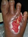
Tissue type within the wound bed[edit|edit source]
组织可以被描述为可行的或non-viable.
- Viable tissueis bright, shiny, bouncy, taut, and moist. Viable tissue can be either granulation tissue or epithelial tissue.
- Granulation tissueis the new growth of small blood vessels and connective tissue. It is soft and spongy, and may bleed when touched. Granulation tissue is usually pale pink initially, and then beefy red as it is vascularised.
- Healthy granulation tissue is bright red, shiny, granular, or bumpy, with a velvety appearance.
- Unhealthy granulation tissue is pale or dull red due to poor vascular supply. It may also be friable and disintegrate with dressing removal or with cleansing.
- Hypergranulation is an overgrowth of granulation tissue that rises above the level of the wound surface. It is irregular in shape and has large or swollen granules. Hypergranulation is typically due to maceration, infection, or friction on the wound bed. It must be removed for proper healing to occur.
- Epithelial tissueis pink or red in colour and has the appearance of new skin.
- Granulation tissueis the new growth of small blood vessels and connective tissue. It is soft and spongy, and may bleed when touched. Granulation tissue is usually pale pink initially, and then beefy red as it is vascularised.
- 对于没有生存能力的组织is necrotic or dead tissue within the wound bed. It must be removed for proper healing to occur and to decrease infection risk.
- Sloughis non-viable subcutaneous tissue and by-products. It's usually yellow to tan in colour and can be mucousy or stringy. It's a result of the body breaking down dead cells and ranges from non-adherent to loosely adherent to tissue.
- Escharindicates deeper tissue damage. It can be black, grey, or brown. It is adherent to the wound bed and may be hard, spongy, rubbery, or leathery. Eschar is sometimes confused with scabs. Ascabis a collection of dried blood cells, platelets, and serum on top of the skin surface, and the healing actually occurs beneath the scab, whereas eschar is non-viable tissue that must be removed to allow healing to occur.[1][3]
-

Healthy beefy granulation tissue.
-
White arrows indicate areas of advancing epithelial cells.
-

White arrows point to examples of hypergranulation tissue.
-

Yellow area denotes slough, pink arrow shows new epithelial cells, white arrow highlights areas of periwound maceration. Differentiating tissue types is important for debridement purposes.
-

Yellow arrow denotes black eschar, white arrow shows macerated periwound, red areas indicate areas of erythema.
-
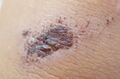
Image of a scab for comparison with eschar.


All images provided by and used with kind permission of Dana Palmer PT.
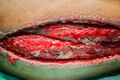
Anatomical structures visible within the wound[edit|edit source]
It's important to document normal anatomical structures that are visible in the wound and to continuously monitor their health and viability. It is vital to be able to recognise tissue types for identification and debridement purposes.
- Blood vessels在健康的时候是紫色的颜色,黑色或br吗own in colour when unhealthy, clogged, or calcified.
- Fatis yellow and globular when healthy and appears shrivelled, brown when unhealthy.
- Muscleis pink or dark red, striated, firm, and resilient to pressure when healthy. It may jump or twitch when probed. When unhealthy, it appears brown, shrivelled, friable, and does not twitch.
- Tendonsare white and shiny when healthy. They are covered with a white tendon sheath. When unhealthy, the sheath will be frayed or stringy, and the tendon will become yellow or brown.Ligaments and joint capsuleshave a similar appearance to tendons when healthy and unhealthy.
- Boneis beige or tan, hard and covered with a clear membrane (the periosteum) when healthy. When unhealthy, it is brown or black, and may be friable and disintegrate with palpation.
Document the percentage of tissue types, colour, and consistency seen in the wound bed.[1]
-

红色箭头指向钙化提单ood vessels.Image used with kind permission of Dana Palmer PT.
-

Yellow arrow indicates fat.
-
-

-

-

White arrow indicates bone, red arrow points to muscle.


Wound drainage[edit|edit source]
When describing drainage (exudate), consider both the type and amount. Make note of drainage prior to removing dressing if there is strike through drainage on the outside of the dressing. Note drainage on the inside of the dressing before cleansing the wound. It is also important to note the amount of time since the last dressing change. Make note of any additional drainage that occurs after wound cleansing and during the wound care interventions.
There are four primary types of wound drainage and two additional types of drainage (serosanguineous and seropurulent) that are combinations of the primary types.
| Drainage Type | Description | When to Refer or Reassess | Example |
|---|---|---|---|
| Sanguineous drainage |
|
|
|
| Serous drainage |
|
If copious, can indicate trauma to the wound, chronic inflammation due to biofilm or localised infection. | |
| Serosanguineous drainage |
|
If serous drainage changes to serosanguineouslaterin the healing process, it may indicate new trauma has occurred to the wound. | |
| Purulent drainage |
|
|
|
| Seropurulent drainage | Occurs when serous drainage starts to turn cloudy, yellow, or tan. |
|
|
| Autolytic drainage |
|


Terminology used to describe amount of drainage:
| Dressing | Wound bed | Drainage during treatment | |
|---|---|---|---|
| None | Dry | Dry | None |
| Scant |
|
Moist | None |
| Minimal |
|
Moist | There may be some drainage on dressing removal, but no drainage occurs during treatment. |
| Moderate |
|
Wet | Some drainage is visible in the wound bed after dressing removal, as well as some drainage occuring during treatment. |
| Heavy |
|
Very wet | Drainage is visible immediately upon dressing removal, and continues throughout treatment. |
| Copious |
|
Filled with fluid and saturated |
|
Wound Odour[edit|edit source]
- Almost all wounds have some odour on dressing removal. Odour does not automatically suggest infection.
- Odour is concerning if the smell lingers after dressing removal and wound cleansing.
- Document the odour of the wound and not the odour from the topical agent, dressing, or the dressing by-product.
- For example: if Dakin's solution was used, there will be a smell like bleach, if Burrow's solution was used, it may smell like vinegar, povidone-iodine (betadine) will smell like iodine. Hydrocolloid dressings will have a distinctive smell as a result of the chemical reaction from autolytic debridement.[1]
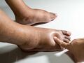
Periwound Skin[edit|edit source]
Periwound skin is the area around a wound that may be affected by "wound-related factors and/or underlying pathology" according to LeBlanc et al.[4]
Periwound assessment should include the following:[1]
- Trophic changes:changes in skin texture, changes in nail beds, loss of hair, shiny skin
- Skin discolouration:
- Haemosiderin depositsare commonly seen in venous insufficiency
- White spots calledatrophie blancheare commonly seen with arterial disease
- Ahaematoma(purple ecchymosis) points to possibility of deep tissue destruction
- You may also see redness, darkening, purple spots, or other pigment changes that should be documented
- Macerationis softening of tissue caused by excessive moisture. This tissue appears white due to a loss of pigmentation, and it is often soft and soggy
- Indurationis abnormal firmness of tissue with margins and sometimes has an orange peel appearance
- Hydration or turgor
- Hyperkeratosis(callus)
- Oedema:
- Delays healing
- 是一个正常的反应炎症pha的e
- Other causes could include: (1) dependent position, (2) venous insufficiency, (3) renal failure, or (4) right-sided congestive heart failure
- Pitting oedemais assessed by pressing your finger into the skin for five seconds and then releasing your finger. If the indentation stays, it's termed pitting oedema. Pitting oedema is often seen in congestive heart failure, venous insufficiency, and with DVTs
- Non-pitting oedemais stretched skin that is shiny and hard and is often seen in lymphoedema or angioedema
-

Haemosiderin staining
-

Atrophie blanche
-

White arrow indicates area of maceration in periwound
-

White arrow indicates area of induration
-
Pitting oedema


Please see the following 1-minute video for a demonstration of the assessment pitting oedema.
Blood flow[edit|edit source]
- Palpate the posterior tibial pulse and the dorsalis pedis pulse, document the presence and quality
- If possible, perform theankle brachial indexand document the result[1]


Sensation[edit|edit source]
- Semmes-Weinstein monofilamentsfor protective sensation[6]
- Light touch and pressure manually
- Heat/cold
- Proprioception[1]
Infection[edit|edit source]
- When assessing for infection, utilise the acronym IFEE: induration, fever, erythema, and (o)edema.
- Key characteristics of infectioninclude (1) streaks of redness, (2) increased warmth, (3) intense pain, (4) significant oedema, (5) exudate changes from serous to purulent, and (6) a strong odour.
- Systemic characteristic of infectioninclude (1) fever greater than 38.3 degrees Celsius (101 degrees Fahrenheit), (2) chills, (3) lethargy, (4) restlessness, and (5) confusion.
- Reddened skin with streaks leading away from the wound may mean cellulitis or necrotising fasciitis, an infection of the surrounding tissues that can be life and limb threatening. Necrotising fasciitis requires immediate emergency medicine consultation.[1]
If an infection is suspected, a swab culture may be ordered. And in most practice environments, a wound care therapist can perform a swab culture.
- Use the Levine technique when performing a swab culture (see additional resources for more information)
- Do not culture eschar, slough, or other non-viable tissue; this will result in a false positive culture from the bacteria present in the non-viable tissue.[1]
Joint Biomechanical Function[edit|edit source]
For the joints proximal and distal to the wound:[1]
- Passive and active range of motion
- Manual muscle testing
- Reflex testing of the involved extremity to assess for a neuropathy
Wound Classification[edit|edit source]
Wounds can be classified in several different categories:[1]
- Acute or chronic.Chronic wounds are wounds that have not finished the proliferative phase at the end of four weeks.
- Wounds are also classified based ondepth, and different wound types have different classification systems.
- One way to classify depth is superficial, superficial partial thickness, deep partial thickness, and full thickness.
- Neuropathic ulcers can be staged usingWagner’s Classification.[7]
- Another common way to classify depth is based on theBates-Jensen Wound Assessment Tool.[8]
- Pressure injuries or pressure ulcers are staged using a system developed by theNational Pressure Injury Advisory Panel.
Common characteristics of leg ulcers for classification[1]
| Common cause | Pain | Common location and appearance | Example | |
|---|---|---|---|---|
| Arterial ulcers | Arteriosclerosis | Very painful |
|
|
| Venous ulcers | Venous insufficiency | Mild |
|
|
| Neuropathic ulcers | Diabetes | None due to neuropathy, however occasionally there will be pain sensations in the limb that is unrelated to the wound. |
|


- To review information about arterial and venous ulcers, please readthis article.
- To review information about neuropathic ulcers, please readthis article.
Resources[edit|edit source]
- 'Please view the following optional 7-minute video for a discussion of the steps of wound swab culture collection via the Levine technique (starts at time 2:40), plus an overview of setup and documentation.
- Wound Culture Collection: Levine Technique Check-list
- Thepitting oedema severity scale
- TheBates-Jensen Wound Assessment Tool
- The National Pressure Injury Advisory PanelPressure Injury Stages
- The National Pressure Injury Advisory PanelPressure Injury Prevention Protocol
References[edit|edit source]
- ↑1.001.011.021.031.041.051.061.071.081.091.101.111.121.131.141.151.161.171.181.191.20Palmer, D. Integumentary Physiotherapy Programme. Basic Wound Assessment. Physioplus. 2023.
- ↑YouTube. Principles of Draping Patients for Physical Exams. Available from:https://www.youtube.com/watch?v=Q6oCdxISRCE&t=85s[last accessed 13 Feb 2023]
- ↑3.03.1Moura CD, Dowsett C, Bain K, Bain M.Advancing practice in holistic wound management: a consensus-based call to action.Wounds International. 2020;11(4):70-5.
- ↑LeBlanc K, Beeckman D, Campbell K et al (2021)Best practice recommendations for prevention and management of periwound skin complications.
- ↑YouTube. Pitting Edema | Vivo Phys. Available from:https://www.youtube.com/watch?v=adkrWQ8sWFU[last accessed 13 Feb 2023]
- ↑Castellano VK, Jackson RL, Zabala ME.Contact mechanics modeling of the Semmes‐Weinstein monofilament on the plantar surface of the foot.Int J脚脚踝。2021;5(2):055。
- ↑Shah P, Inturi R, Anne D, Jadhav D, Viswambharan V, Khadilkar R, Dnyanmote A, Shahi S.Wagner's classification as a tool for treating diabetic foot ulcers: Our observations at a suburban teaching hospital.Cureus. 2022 Jan 22;14(1).
- ↑Bates‐Jensen BM, McCreath HE, Harputlu D, Patlan A.Reliability of the Bates‐Jensen wound assessment tool for pressure injury assessment: The pressure ulcer detection study.Wound Repair and Regeneration. 2019 Jul;27(4):386-95.
- ↑YouTube. Wound Culture | Wound swab for culture and sensitivity | How to collect wound culture. Available from:https://www.youtube.com/watch?v=zDuxe0AH3Ac[last accessed 07/March/2023]

