
Ankle and Foot
Anatomy[edit|edit source]
Theankleis the part of the lower limb encompassing the distal portion of the leg and proximal portions of the foot. Theankleencompasses theankle joint, an articulation between thetibiaandfibulaof the leg and thetalusof thefoot. See the page forankle jointfor more information.
Thefootis the part of the lower limb distal to theankle joint. It is covered on its dorsal surface by loosely adherent skin and on its plantar/inferior surface by thick hairless skin that is tough and strongly adherent to the underlying plantar aponeurosis. Thefootcontains 26 small bones that are designed for weight bearing and force distribution. The bony alignment creates three arches the provide efficient weight distribution while avoiding compression of plantar neuro-vascular structures. The three arches,medial and lateral longitudinal and the transverse archtogether create an architectural vault, which is one of the strongest load-bearing structures known to mankind.[1]
Bones[edit|edit source]
The bones of the foot are named as follows:

- The metatarsals - numbered from medial or first (big toe), to lateral or fifth (little toe)
- The phalanges - toes 2-5 each have 3 phalanges. The first or big toe (hallux) has only two[2]
Muscles[edit|edit source]

The dorsum of thefoot只有一个肌肉(可能2根据classification). This is the extensor digitorum brevis (some authors name the most medial part of this muscle extensor hallucis brevis). Tendons are the main collagenous structures in the dorsum. The tendons connect anterior/dorsiflexor compartment muscles of the leg to the foot bones.
The plantar aspect of the foot contains the tough fibrous plantar aponeurosis covering muscles and tendons arranged in 4 layers, numbered from 1 superficial to 4 deep:
- 第一层由of theabductor didgiti minimi, flexor digitorum brevis, abductor hallucis
- Layer 2 consists of thequadratus plantae, the lumbricals, and the long tendons offlexor digitorum longusandflexor hallucis longus
- Layer 3 consists of theflexor hallucis brevis,adductor hallucisand屈肌digiti最小的短
- Layer 4 consists of the interosseous muscles and the long tendons ofperoneus/fibularis longusandtibialis posterior[3]
Ligaments[edit|edit source]
Ankleligament injury is the most frequent cause of acute ankle pain. Hence, it is important to understand the anatomy of ankle ligaments for correct diagnosis and treatment.
The ligaments around the ankle can be divided, depending on their anatomic position, into three groups: the lateral ligaments, the deltoid ligament on the medial side, and the ligaments of the tibiofibular syndesmosis that join the distal epiphyses of the tibia and fibula.[4]
Thelateral collateral ligament complex (LCL)consists of[4]:

- Anterior talofibular ligament: it is the most frequently injured ligament of theankle. This ligament plays an important role in limiting anterior displacement of thetalusand plantar flexion of theankle.
- Posterior talofibular ligament: The posterior talofibular ligament originates from the malleolar fossa, located on the medial surface of the lateral malleolus, coursing almost horizontally to insert in the posterolateral talus. It is the strongest ligament of the lateral ankle. plays only a supplementary role in ankle stability when the lateral ligament complex is intact.
- Calcaneofibular ligament: The calcaneofibular ligament originates from the anterior part of the lateral malleolus. Its primary role is to restrain inversion in a neutral or dorsiflexed position, restrains subtalar inversion, thereby limiting talar tilt within the ankle mortise.
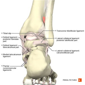

The内侧副韧带(制程), also known as deltoid ligament, is composed of two layers; superficial and deep. The MCL is a multifascicular ligament, originating from the medial malleolus to insert in thetalus,calcaneus, and navicular bone. It primary restrains to valgus tilting of thetalus. Both the superficial and deep layers individually resist eversion of the hindfoot. It also stabilizesankleagainst plantar flexion, external rotation, and pronation.[4]
The ligaments of the tibiofibular syndesmosis consist of anterior or anteroinferior tibiofibular ligament, the posterior or posteroinferior tibiofibular ligament, and the interosseous tibiofibular ligament. The syndesmotic ligament complex ensures the stability between the distaltibiaand thefibulaand resists the axial, rotational, and translational forces that attempt to separate thetibiaandfibula.[4]
Neurovasculature[edit|edit source]

.jpg)
Cutaneous innervation of the dorsum is by the superficial and deep peroneal/fibular nerves. Cutaneous innervation of the plantar aspect is by the medial and lateral plantar and tibial nerves. Dorsal motor innervation is by the deep peroneal nerve to extensor digitorum brevis and extensor hallucis brevis. Plantar motor innervation is via the medial and lateral plantar nerves (terminal branches of the tibial nerve). Arteries crossing into the foot accompany nerves of corresponding names. Therefore the anterior tibial ordorsalis pedis artery, and the posterior tibial artery, are the terminal branches of the medial and lateral plantar arteries.[5]
Clinical Examination[edit|edit source]
Ankle & Foot Examination[edit|edit source]
Special Tests[edit|edit source]
Outcome Measures[edit|edit source]
Conditions[edit|edit source]
- Achilles tendonitis
- Achilles rupture
- Ankle & foot fractures
- Ankle & foot arthropathies
- Ankle sprain
- Ankle Impingement
- Anterior ankle impingement syndrome
- Ankle ssteochondral lesions
- Calcaneal fractures
- Calcaneal spurs
- Compartment syndrome of the foot
- Compartment syndrome of the lower leg
- Calf strain
- Hallux valgus
- Hallux rigiditus
- Lisfranc injuries
- Metatarsalgia
- Peroneal rendonitis
- Plantarfasciitis
- Posterior tibial tendon dysfunction
- Retrocalcaneal bursitis
- Shin splints
- Sinus tarsi syndrome
- Tarsal tunnel syndrome
- Tibiofibular diastasis[14]
Procedures[edit|edit source]
References[edit|edit source]
- ↑The Editors of Encyclopaedia Britannica. Foot. Available from:https://www.britannica.com/science/foot(accessed 26/02/2019).
- ↑Schmidler C. Anatomy of the Foot and Ankle & Common Problems. Available from:https://www.healthpages.org/anatomy-function/anatomy-foot-ankle/(accessed: 25/02/2019)
- ↑Arthritis Foundation. Anatomy of the foot. Available from:https://www.arthritis/where-it-hurts/foot-heel-and-toe-pain/foot-anatomy.php(accessed 25/02/2019).
- ↑4.04.14.24.3Golanó P, Vega J, De Leeuw PA, Malagelada F, Manzanares MC, Götzens V, Van Dijk CN.Anatomy of the ankle ligaments: a pictorial essay.Knee Surgery, Sports Traumatology, Arthroscopy 2010;18(5):557-69.
- ↑Hernández-Díaz C, Saavedra MÁ, Navarro-Zarza JE, Canoso JJ, Villasenor-Ovies P, Vargas A, Kalish RA.Clinical anatomy of the ankle and foot.Reumatologia clinica 2012;8:46-52.
- ↑CRTecnologies. Anterior Drawer Test Ankle. Available from:https://www.youtube.com/watch?v=Z4rvAT3a7OY[last accessed 30/6/2021]
- ↑bigesor. Talar Tilt Test. Available from:https://www.youtube.com/watch?v=2qF_DOe2jPE[last accessed 30/6/2021]
- ↑Physical Therapy Nation.Syndesmosis Squeeze Test. Available from:https://www.youtube.com/watch?v=rM9Rk1oucHM[last accessed 30/6/2021]
- ↑Daryl Lawson. Windlass Test Nonweightbearing. Available from:https://www.youtube.com/watch?v=iQD5qtO5-zE[last accessed 30/6/2021]
- ↑Kate Cornet. Windlass Test. Available from:https://www.youtube.com/watch?v=ZO0wREhjxH0[last accessed 30/6/2021]
- ↑Alazzawi S, Sukeik M, King D, Vemulapalli K.Foot and ankle history and clinical examination: A guide to everyday practice.World Journal of Orthopedics 2017;8(1):21–29.
- ↑Fraser JJ, Koldenhoven RM, Saliba SA, Hertel J.Reliability of ankle-foot morphology, mobility, strength, and motor performance measures.International Journal of Sports Physical Therapy 2017 Dec;12(7):1134.
- ↑Martin RL, Davenport TE, Reischl SF, McPoil TG, Matheson JW, Wukich DK, McDonough CM, Altman RD, Beattie P, Cornwall M, Davis I.Heel pain—plantar fasciitis: revision 2014.Journal of Orthopaedic & Sports Physical Therapy 2014;44(11):A1-33.
- ↑American College of Foot and Ankle Surgeons. Browse Foot & Ankle Conditions. Available from:https://www.foothealthfacts.org/foot-ankle-conditions/browse-foot-ankle-conditions(accessed 25/02/2019).
- ↑Coster C.D, Bradly J, Solorzano J, Buxton S, Williams D. Total Ankle Arthroplasty. Available from://m.houseofhawgs.com/Total_Ankle_Arthroplasty(accessed 25.02.2019)

